
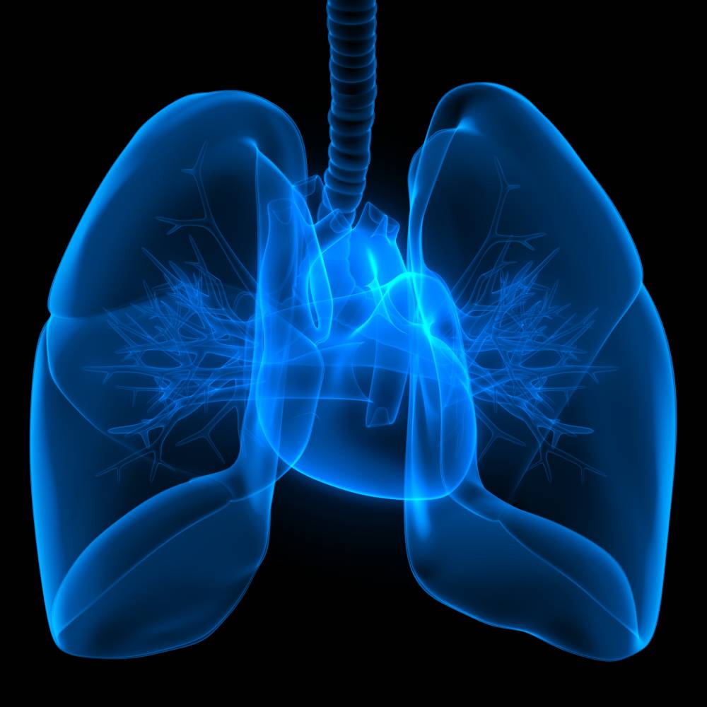
Atelectasis in Anesthesia: Causes and Management
The term “atelectasis” originates from the Greek words atelez and ektasiz, meaning “imperfect” and “expansion” respectively,1 and describes an incomplete expansion of lung tissue, resulting from the partial or complete collapse of the small airways and leading to an impaired exchange of CO2 and O2.1,2 A study in 1995 estimated that the incidence of atelectasis in patients undergoing general anesthesia is around 90%.1 This condition typically occurs within 72 hours of general anesthesia and is a common postoperative complication.1
Atelectasis can be caused by compression of lung tissue (compressive), absorption of alveolar air (resorptive), or impaired pulmonary surfactant production.3 Compression atelectasis is the result of a decreased transmural pressure gradient across the alveolus, leading to alveolar collapse.1,3 Resorptive atelectasis occurs when alveolar air gets absorbed distal to an obstructive lesion.1,3 The obstruction either partially or completely inhibits ventilation to the area.1 Over time, all of the air in that segment will be absorbed and, without return of ventilation, the airway will collapse.1,3 Children are especially susceptible to the resorption form because they have poorly developed collateral pathways for ventilation.1
Roughly 15 to 20% of the lung at its base can collapse during uneventful anesthesia before any surgical intervention.1 Research has shown that atelectasis can appear in the dependent regions of both lungs within five minutes of induction of anesthesia.4 This complication does not preferentially affect men or women, and there is no increased incidence in patients with asthma or increased age.1,5 Both obese and pregnant patients have an increased risk of atelectasis due to decreased functional residual capacity and compliance.6 Atelectasis appears more frequent after cardiac surgery with cardio-pulmonary bypass than after other types of surgery.1 Inadequate pain control can also contribute to its development by causing shallow breathing or by preventing coughing.1
Often, atelectasis is asymptomatic. However, patients may present with decreased or absent breath sounds, cough, sputum production, tachypnea, or diminished chest expansion.1 A chest x-ray, chest CT, and fiberoptic bronchoscopy are useful for diagnosing this complication.1 A chest x-ray will depict atelectasis as the displacement of interlobular fissures, pulmonary opacification, or tracheal shift toward the affected side.1 Chest CT commonly reveals dependent lung densities and the loss of volume in the affected side of the chest.1 Fiberoptic bronchoscopy can be used as both a diagnostic and therapeutic tool, especially for revealing the cause of any contributing obstruction .1
The majority of atelectasis cases that appear during general anesthesia lead to transient lung dysfunction that quickly resolves itself.1 However, some patients develop significant perioperative respiratory complications that can lead to increased morbidity and mortality if not treated.1 The use of continuous positive airway pressure and positive end-expiratory pressure (PEEP) help to prevent the development of atelectasis.8 Other methods that have been shown to decrease incidence involve changing the patient’s position from supine to upright, encouraging the patient to take deep breaths, chest physiotherapy, and tracheal suctioning.7,8 Pharmacologic treatment options include mucolytic agents (acetylcysteine) and recombinant human DNase.1 As mentioned previously, fiberoptic bronchoscopy also has a role in the management of atelectasis.1 One study showed that fiberoptic bronchoscopy led to improved lung function and reversal of atelectasis in 76% of cases.9
References
- Grott, K., & Dunlap, J. (2020). Atelectasis. In StatPearls [Internet]. StatPearls Publishing LLC. Retrieved 29 September 2020, from https://www.ncbi.nlm.nih.gov/books/NBK545316/
- Carlsen, K., Crowley, S., & Smevik, B. (2019). Atelectasis. In R. Wilmott, A. Li, P. Sly, A. Bush, R. Deterding, F. Ratjen & H. Zar, Kendig’s Disorders of the Respiratory Tract in Children (9th ed.). Elsevier Inc. Retrieved 29 September 2020, from https://doi.org/10.1016/C2015-0-01292-8
- Peroni, D., & Boner, A. (2000). Atelectasis: mechanisms, diagnosis and management. Paediatric Respiratory Reviews, 1(3), 274-278. https://doi.org/10.1053/prrv.2000.0059
- Brismar, B., Hedenstierna, G., Lundquist, H., Strandberg, Å., Svensson, L., & Tokics, L. (1985). Pulmonary Densities during Anesthesia with Muscular Relaxation—A Proposal of Atelectasis. Anesthesiology, 62(4), 422-428. https://doi.org/10.1097/00000542-198504000-00009
- Gunnarsson, L., Tokics, L., Gustavsson, H., & Hedenstierna, G. (1991). Influence of Age on Atelectasis Formation and Gas Exchange Impairment During General Anaesthesia. British Journal of Anaesthesia, 66(4), 423-432. https://doi.org/10.1093/bja/66.4.423
- Coussa, M., Proietti, S., Schnyder, P., Frascarolo, P., Suter, M., Spahn, D., & Magnusson, L. (2004). Prevention of Atelectasis Formation During the Induction of General Anesthesia in Morbidly Obese Patients. Anesthesia & Analgesia, 1491-1495. https://doi.org/10.1213/01.ane.0000111743.61132.99
- Toshida, K., Minagawa, R., Kayashima, H., Yoshiya, S., Koga, T., & Kajiyama, K. et al. (2020). The Effect of Prone Positioning as Postoperative Physiotherapy to Prevent Atelectasis After Hepatectomy. World Journal of Surgery. https://doi.org/10.1007/s00268-020-05682-0
- Östberg, E., Auner, U., Enlund, M., Zetterström, H., & Edmark, L. (2017). Minimizing atelectasis formation during general anaesthesia—oxygen washout is a non-essential supplement to PEEP. Upsala Journal of Medical Sciences, 122(2), 92-98. https://doi.org/10.1080/03009734.2017.1294635
- Restrepo, R., & Braverman, J. (2014). Current challenges in the recognition, prevention and treatment of perioperative pulmonary atelectasis. Expert Review of Respiratory Medicine, 9(1), 97-107. https://doi.org/10.1586/17476348.2015.996134